X-ray Pulse Duration and Anatomical Noise Studies at ECR 2015
Reading Time: 3 minutes read
In March, I will be presenting two papers at the European Society of Radiology’s 2015 European Congress of Radiology. Both papers are in collaboration with Dr. Narinder Paul, Division Chief of Cardiothoracic Radiology and his group at the University Health Network in Toronto, Canada. It is a pleasure to be able to report our team’s studies that take a look at lung nodules, and examine them in two ways:
- Measuring the influence of X-ray pulse duration on the module during digital radiography (DR) in a dynamic anthropomorphic chest phantom
- Observing anatomical noise and its impact on lung nodule conspicuity by comparing DR, dual-energy X-ray, and digital angular tomosynthesis (3D imaging)
The purpose behind the first paper was to observe the impact of cardiac motion on lung nodule margin. Lung nodule margin is an important component to the evaluation of interval change on surveillance imaging. Patients that are breathless or anxious may have increased cardiac motion which may impact nodule definition. As a result, we investigated whether shortening the X-ray pulse duration reduces the impact of cardiac motion on the sharpness of nodule margins for digital radiography.
The method and materials we used to collect our data included an anthropomorphic phantom that was modified to simulate breathing and cardiac motion. Two spherical nodules were placed in the left lung and images were acquired under a range of pulses while adjusting the mA to keep constant mAs. The cardiac rate was set to consistent rates and the exam was repeated 10 times for each ms setting. Ultimately, an algorithm was developed to locate the nodules and derive an average radial profile to yield a sharpness metric generated from the slope of the profile.
From this study, we discovered that pulse duration can impact lung nodule sharpness due to cardiac motion. As a result, care should be taken when assigning protocols for nodule screening.
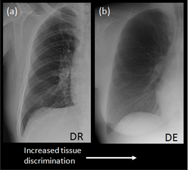
Illustration of tissue discrimination in x-ray medical imaging resulting in reduced background anatomical noise and improved feature conspicuity. DR – Digital radiography, DE – Dual-energy x-ray image.
The second paper that I will be presenting live at ECR, we will be focusing on background anatomical noise as it is a leading cause of reduced object conspicuity in digital radiographic (DR) imaging. Dual-energy (DE) imaging reduces the influence of anatomical noise via tissue discrimination, while digital tomosynthesis (DT) reduces anatomical noise via depth discrimination.
Our methods to the study involved an anthropomorphic chest phantom with a spherical nodule that was imaged with DR, DE, and DT with the same in-room system. DE images were acquired with fixed and differential filtration and DT images were acquired at three dose levels, while a chest DR provided a performance reference.
The study provided a method for comparing performance across X-ray modalities and some insight in terms of optimal acquisition setting for improved image quality and reduced x-ray dose.
The rest of the results for the study, “Anatomical noise and impact on lung nodule conspicuity: comparing digital radiography, dual-energy X-ray, and digital angular tomosynthesis,” will be shared on Sunday, March 8, as part of the Novel Digital Imaging Techniques session, scheduled between 10:30 – 12:00 at ECR 2015.
You can visit Carestream at ECR 2015 in Booth 210, in Expo B.
Sam Richard, PhD, Senior Research Scientist, Carestream